We’ve probably all heard the phrase “No hoof, no horse” but perhaps never really thought much of it - did you know that the horse’s hoof contains complex structures which scientists are still trying to understand today!?
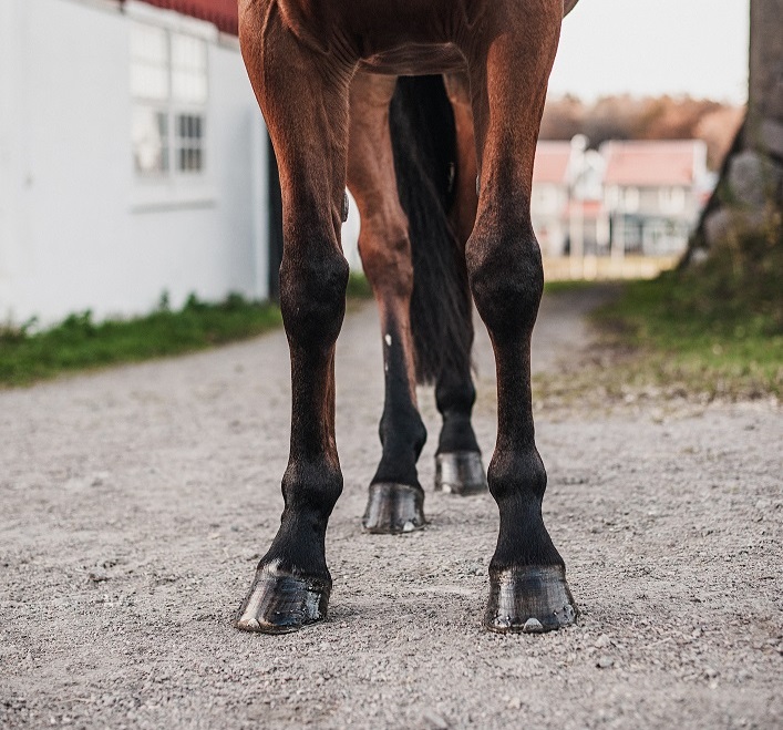
Internal Structure of the Equine Hoof...
The skeletal structure within the hoof contains only the pedal/coffin bone (third phalanx). You will also find the navicular bone and the navicular bursa – a fluid filled sac which helps reduce friction between the deep digital flexor tendon and the bone. The lateral cartilage sits either side of the navicular joint and part of the second phalanx. The cartilage acts as a cushion for the horse’s heel and also helps to support the hoof wall. Between the lateral cartilage we can also find the digital cushion, which expands and flattens when the horse places weight onto that limb.
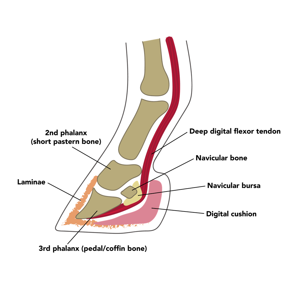
The pedal bone is held in place by a complex arrangement of Laminae. These delicate leaf-like structures attach the pedal bone to the hoof wall. Their structure allows them to hold large amounts of weight while also allowing flexibility of the hoof.
The base of the foot is something we are all familiar with. The hard structure pointing towards the centre of the hoof is known as the frog. It is primarily used as a shock absorber but also plays a role in the circulatory system. Either side of the point of the frog, you will see two groves, these are known as the bars, these work alongside the hoof wall to provide support during weight-bearing.
The inner layer of the hoof wall is known as the white line. It is a pale structure which appears soft and fibrous to the touch. The white line is actually where those laminae attachments have grown out.
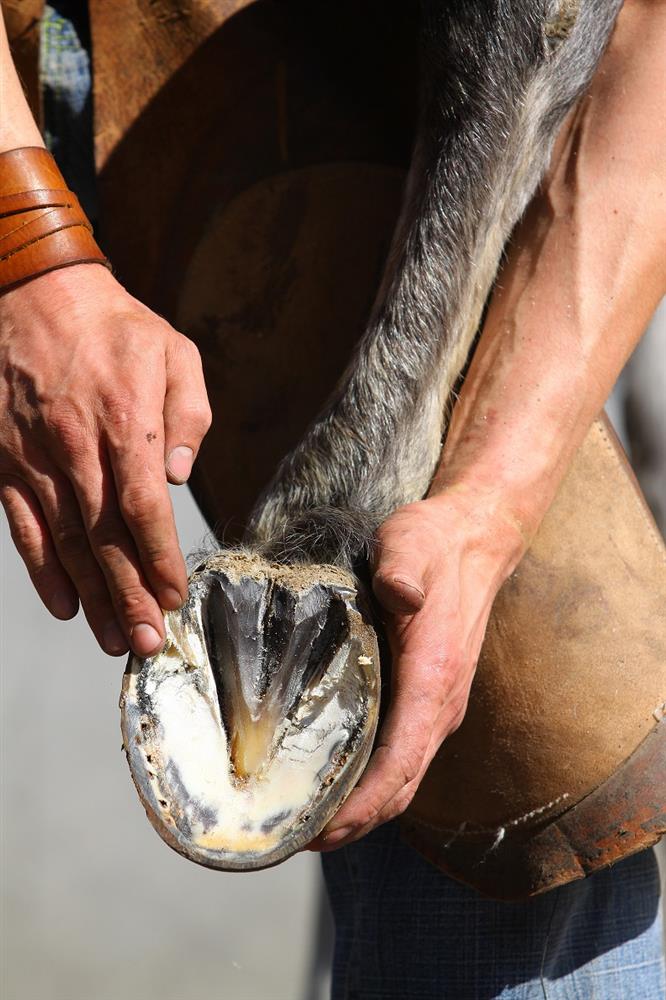
The sole covers the main area of the hoof and is often a grey/white colour. The texture may vary, a horse with shoes on will often have a sole which can easily be scratched at with a hoof pick, whereas a barefoot horse will have a hard sole.
Problems Associated with the Hoof...
Sidebone – Side bone is believed to be caused by excessive concussive forces being applied to the lateral cartilage, repetitive trauma causes the cartilage to harden and become bone-like. This can reduce mobility and can sometime fracture, causing sudden lameness.
Osteoarthritis – The joint between the second and third phalanx is known as the interphalangeal joint. It is almost constantly weight-bearing, which means that it can be at an increased risk of osteoarthritis. Osteoarthritis is often associated with age or trauma to the joint.
Laminitis - This disease is something that horse owners are extremely familiar with and is a constant worry throughout the year! Laminitis is caused by the disruption of blood flow to the laminae causing an inflammatory response. There are a number of causes for this, with the most common being a sudden increase of sugar in the diet. This may be in the form of grains or the horse consuming excessive amounts of lush grass before the horse’s body has a chance to adapt. Laminitis is often linked to underlying conditions such as Cushing’s Disease or Equine Metabolic Syndrome (EMS).
Navicular Syndrome – The name Navicular Syndrome describes the degeneration of different structures associated with the navicular joint. Navicular Syndrome is a vast topic because it can involve many different tendons and ligaments, further information can be found here.
The hoof as a whole is a remarkable structure, evolved to allow horses to run efficiently and fast. It may look rigid, but it twists and flexes with each step the horse takes. If you’re looking for a supplement to support this impressive structure, then take a look at Feedmark’s Hardy Hoof.

